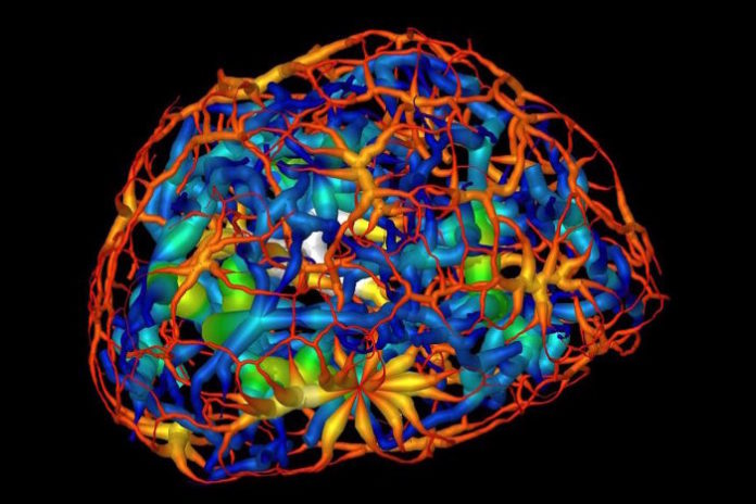
Researchers used powerful X-ray microscope at Berkeley Lab’s Advanced Light Source (ALS) to study differentiating neurons as they mature. Using X-rays, images were taken from many angles to create a three-dimensional composite image from two-dimensional images, showing the changing structure of chromatin during the maturation of olfactory neurons of mice.
Chromatin functions to package DNA so that it can fit inside of a cell, and forms chromosomes. In the video above, take a trip through the nucleus of a cell; in the video below, DNA reorganization in the nucleus a mouse cell is illustrated.
The investigators were also able to take measurements of the packing inside of a type of chromatin, heterochromatin, and revealed more about the role of a protein that aids in the packaging of heterochromatin, and its localization to the nucleus.
“It’s a new way of looking at the nucleus where we don’t have to chemically treat the cell,” explained Carolyn Larabell, the Director of the National Center for X-ray Tomography (NCXT), a collaboration of Berkeley Lab and UC San Francisco (UCSF). “Being able to directly image and quantify changes in the nucleus is enormously important and has been on cell biologists’ wish list for many years.”
She added that Chromatin is “notoriously sensitive” to chemical stains and additives frequently used in biological imaging protocols that highlight regions of interest in a sample. “Until now, it has only been possible to image the nucleus indirectly by staining it, in which case the researcher has to take a leap of faith that the stain was evenly distributed.”
Larabell, who is also a faculty scientist at Berkeley Lab and a UCSF professor, explained that it had been thought that chromatin existed as a group of disconnected islands, but the latest report indicated that chromatin is compartmentalized into two areas of “crowding” that comprise a continuous network throughout the nucleus.
Source: LabRoots


